Visualizing Science Contest Winners for 2026 Span Disciplines
Top-prize image highlights a vital player in the central nervous system.
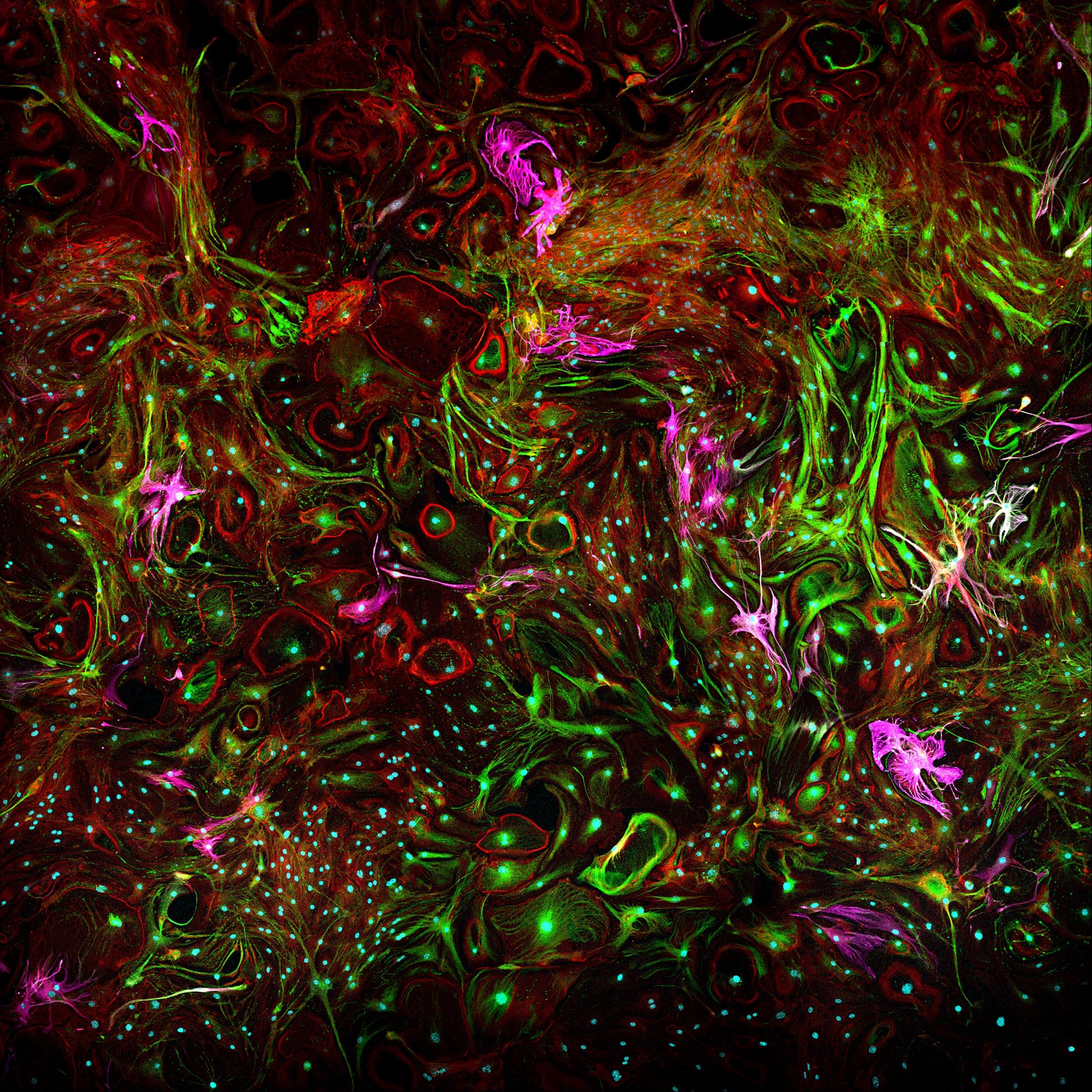
Top Prize Editor’s Choice by Jesse Plotkin, Department of Neuroscience and Waggoner Center for Alcohol and Addiction Research
Every year, the College of Natural Sciences invites faculty, staff and students to send in the most striking and fascinating images from their research for the college-wide Visualizing Science competition. As always, our college community stepped up and delivered.
Jesse Plotkin, a researcher in the Department of Neuroscience and Waggoner Center for Alcohol and Addiction Research, was awarded top honors this year for an eye-catching submission reminiscent of modern art. Because the most effective scientific images grab the viewer, playing on their curiosity to know more, we highlight winning images each year throughout the college-organized Texas Science Festival, displaying work that not only pleases the eye but elevates the mind.
The eight exceptional submissions from the Natural Sciences community that were picked as finalists this year were selected for their beauty and scientific merit by professional science communicators and community members on social media.
All of the images on this page can also be found in our Visualizing Science Showcase, a 3D virtual gallery exhibition which includes winners and finalists from more than a decade of past competitions. A few images in the gallery contain expanded audio descriptions by their creators, including two from this year’s winners.
About the Top Prize Image
Plotkin and other researchers are conducting a study investigating how star-shaped cells called astrocytes, found in the brain and spinal cord and that are responsible for vital functions in the central nervous system, change over time and whether alcohol exposure has an impact on their development.
The cells pictured here, using confocal microscopy, were from a control sample cultured for two weeks under normal conditions. They were stained using immunohistochemistry with antibodies against s100b (green), a calcium-binding protein expressed predominantly in astrocytes, Glial Fibrillary Acidic Protein (magenta), and Phalloidin (red), a fluorescent tag that binds actin. GFAP and actin are proteins found within the structural cytoskeleton of the cells. The entire image was stitched together by software from many smaller images.
Additional 2026 Winners
Dylan Snider, Chemistry Graduate Program
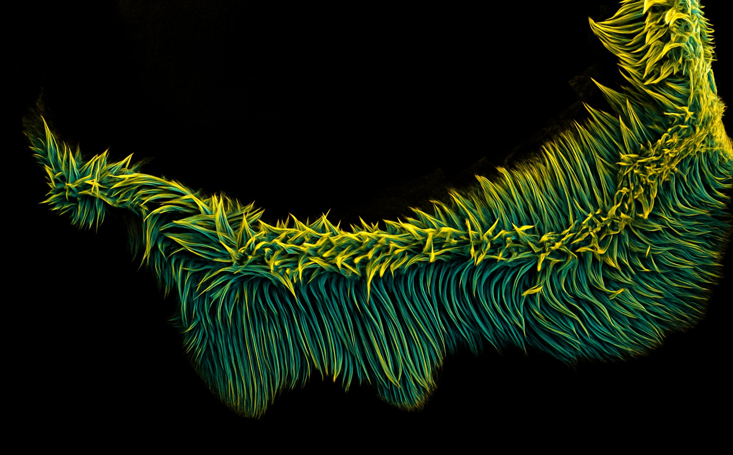
Chemistry Ph.D. candidate Dylan Snider is working on a way to make cancer therapeutics by forcing peptides, short chains of amino acids, to form into small nanofibers around cancer cells by using DNA nanotechnology. While investigating how the peptides reacted without the DNA, he let one of his scanning electron microscope samples dry and found something unexpected on the edge of the sample plate. Instead of the small fibrils or nanofibers he was expecting, Snider found that a large conglomeration of the amino acid chains had condensed into a massive, ethereal, tendril-like structure that could easily be envisioned roaming about on an alien world or even in the depths of our own oceans.
Myles Joyce, Interdisciplinary Neuroscience Graduate Program, Lab of Kristen Harris, Center for Learning and Memory
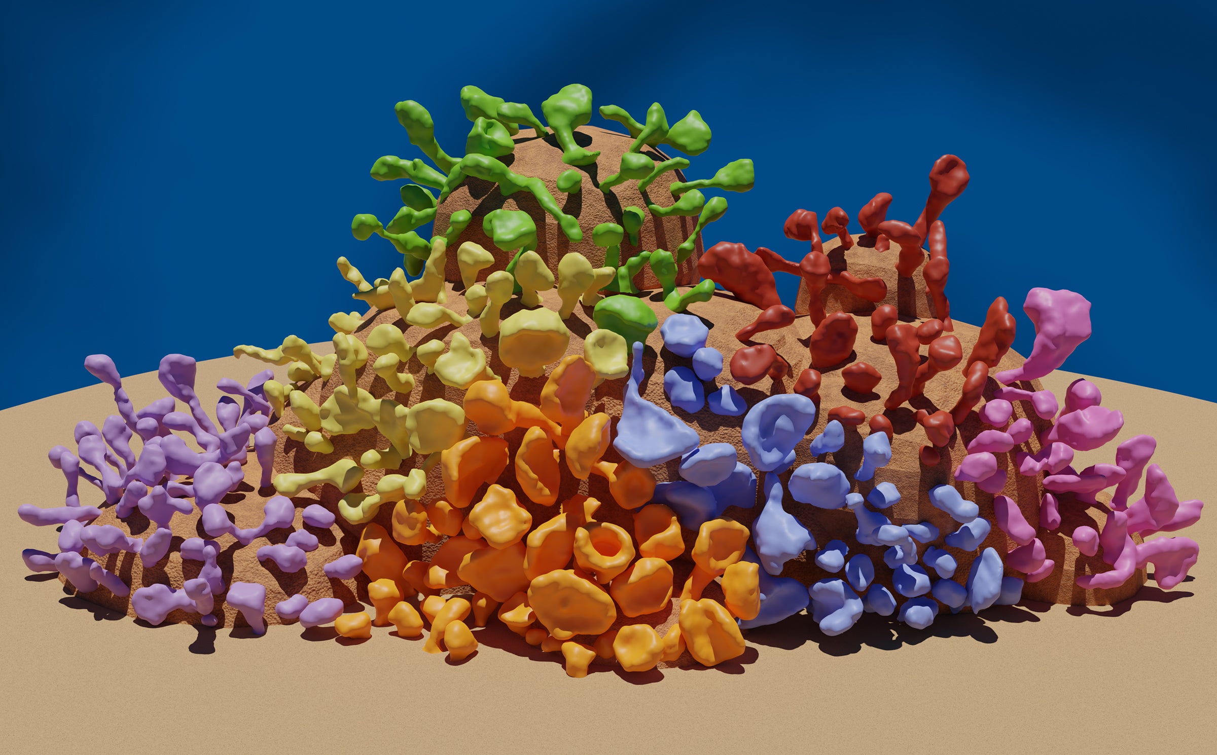
Dendritic spines are specialized protrusions found on the branches of neurons that serve as the main post-synaptic sites for receiving chemical signals in the brain. Highly diverse and dynamic, they change shape throughout life in response to experience and learning. Through countless hours reconstructing rat hippocampal dendrites for his research, neuroscience Ph.D. student Myles Joyce observed how the variety and unique beauty of dendritic spines closely resemble the vibrant biodiversity of coral reefs. In this visual metaphor, each coral polyp is a separate dendritic spine reconstructed from electron micrographs of rat hippocampal neurons. Spines are grouped based on morphology, with different shapes assigned a unique color, highlighting the diversity and complexity of neuronal structure. Just as reefs are built by thousands of distinct yet interdependent organisms, neural connectivity and function arise from the interplay between an orchestra of specialized spines. Nature has a striking tendency to use similar forms and organizational themes across disparate biological systems.
Varanasi Sai Subhankar, Aerospace Engineering Graduate Program and Collaborator with UT Chemistry Department
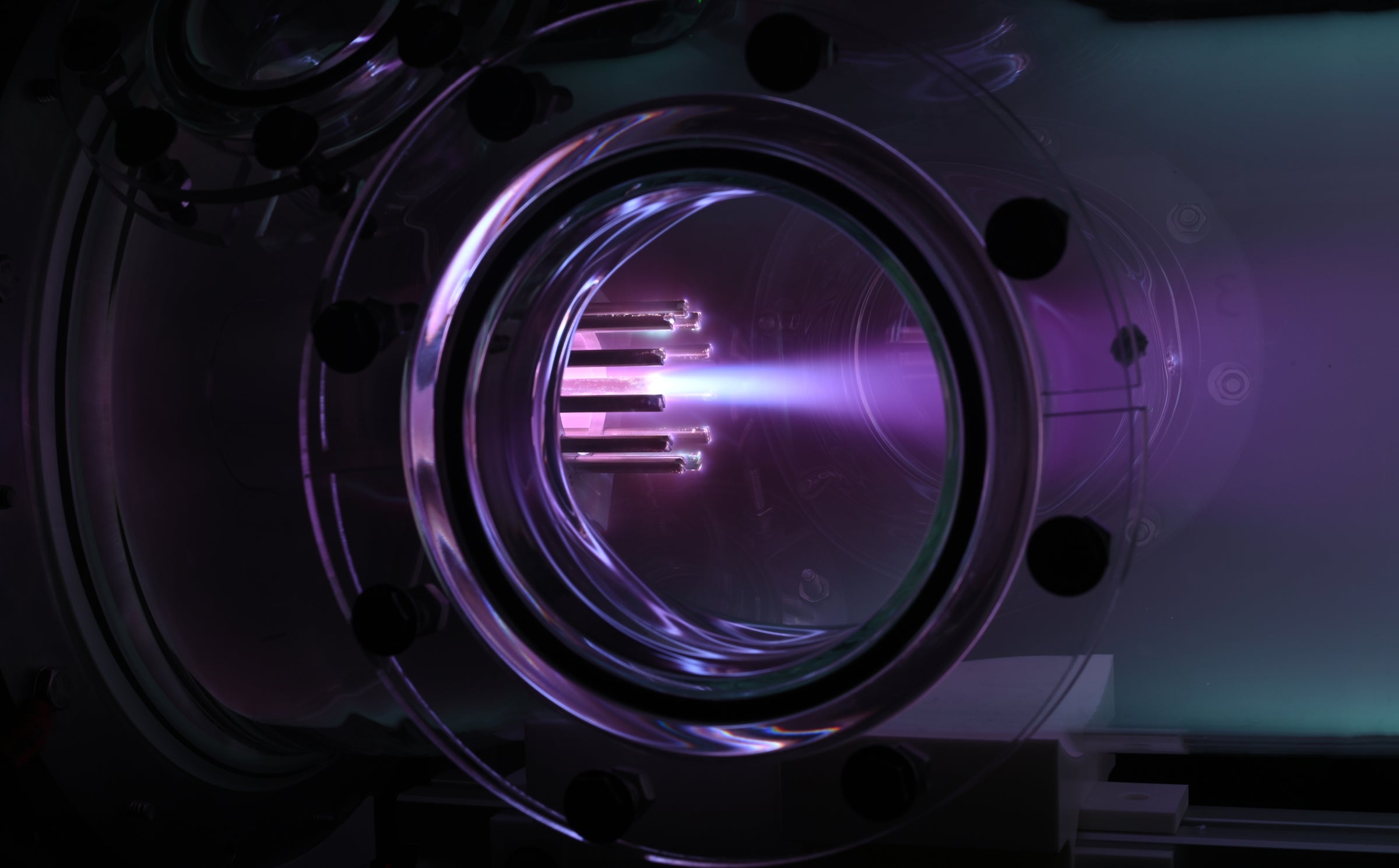
A one-second exposure of the Air-Breathing Deflagration Thruster operating in vacuum shows a bright, collimated plasma jet. Electric current flows through the plasma and interacts with its own magnetic field, generating Lorentz forces that accelerate the plasma outward and simultaneously pinch it inward. The magnetic pinch compresses and heats the plasma before it expands into the vacuum, forming this well-defined jet. The thruster was used to do a variety of experiments to demonstrate the feasibility of air-breathing electric propulsion in Very Low Earth Orbits.
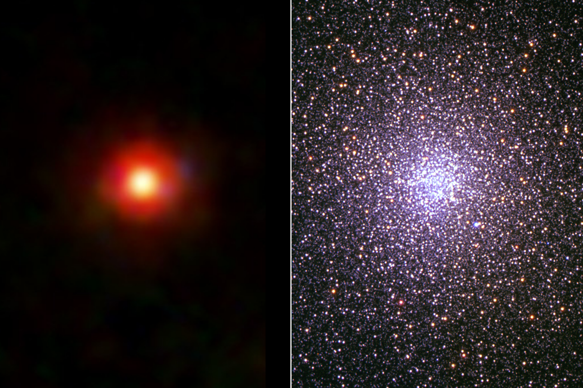
Joohyun Lee, Astronomy Graduate Program, and Paul Shapiro, Department of Astronomy. The image was created using data supplied by Pierre Ocvirk (Strasbourg) and Joe Lewis (Institut d’Astrophysique de Paris) from the CoDa III simulation by Shapiro’s international collaboration, The Cosmic Dawn (“CoDa”) Project, under DOE INCITE AST031 on Summit supercomputer at Oak Ridge National Laboratory, with support from NASA 80NSSC22K1756, image processed at UT TACC under NSF XSEDE TG-AST0900005.
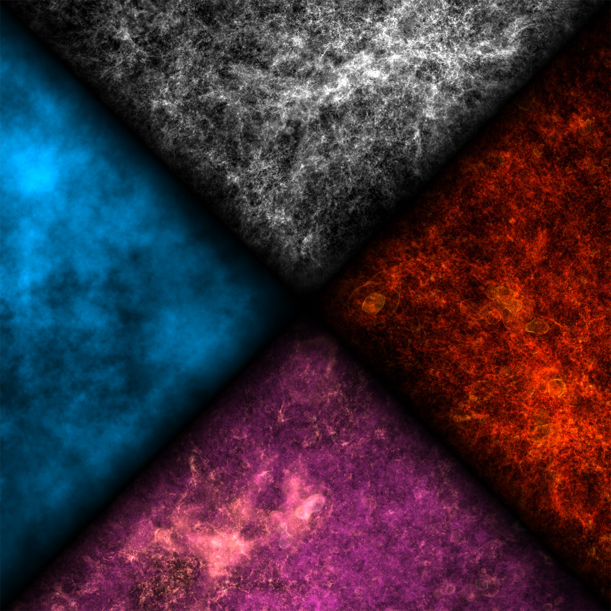
Over time, gravity amplified tiny fluctuations in the featureless ocean of dark and atomic matter that filled the early universe, causing a cosmic web of structure to form, punctuated by galaxies that ionized and heated the intergalactic gas with UV starlight. This evolution was simulated in 3D with hydrodynamics, radiation and gravity, with a trillion computational elements, in a cubic volume 300 million light-years across today. A volume-rendered snapshot when the universe was 770 million years old and 50% ionized, shows the same half-volume in four quantities, rotated clockwise from the top: dark matter (white), heavy elements (red), gas temperature (pink = photo-heated, white = supernova shock-heated, purple = cold) and ionized fraction (blue = ionized, black = neutral). Viewing panels side-by-side reveals an interesting phenomenon: high concentrations of dark matter—reflecting “cosmic variance” in initial conditions—also have high concentrations of other components. Over-dense regions formed galaxies and stars faster than average, causing more ionization, heating and heavy-element enrichment.

Arielle Woznica, Department of Molecular Biosciences
Choanoflagellates are the closest relatives of animals, and these microscopic organisms (each cell is only 5 microns!) help illuminate how we first established symbiotic interactions with bacteria. In this image, which uses super-resolution microscopy to highlight a choanoflagellate colony and its symbiotic bacteria, magenta highlights tubulin, which provides structure to the cell body and the whip-like flagellum that is used for movement and feeding. Green reveals the actin feeding collar, and yellow shows an extracellular structure which connects cells in the colony. Bacteria inside and outside the colony are blue.
Facebook Favorites
Ansar Ali, Department of Molecular Biosciences
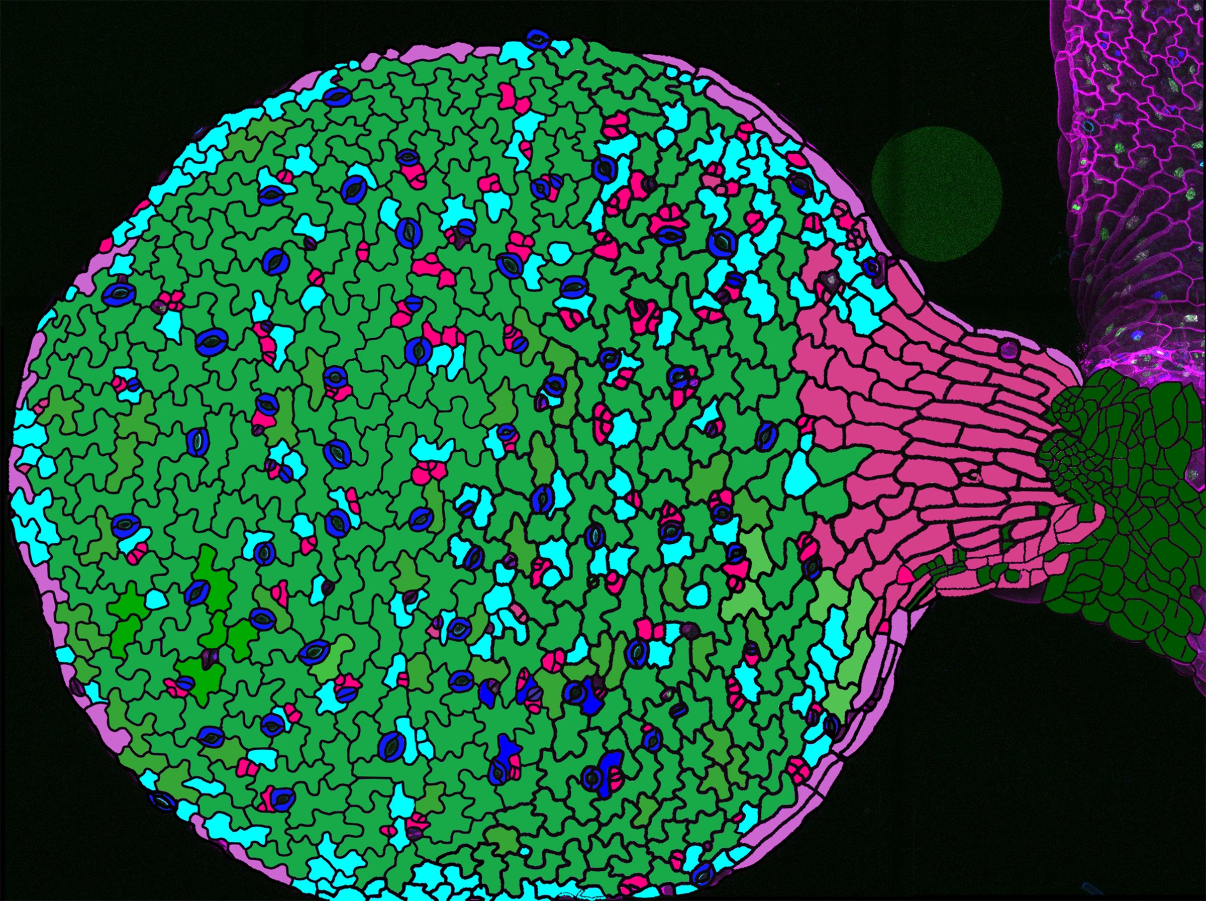
The interlocking pavement cells and stomata on the surface of a leaf create a living mosaic—cell shape, division and fate woven into the plant’s breathable skin. Postdoctoral fellow Ansar Ali created this sketch from a confocal microscopy image of an Arabidopsis leaf that expressed markers for plasma membranes, the lipid bilayers that separate the cell from the outside world. Visible are the jigsaw-like pavement cells (green) that act as armor and the round stomata (blue) that allow the plant to breathe.
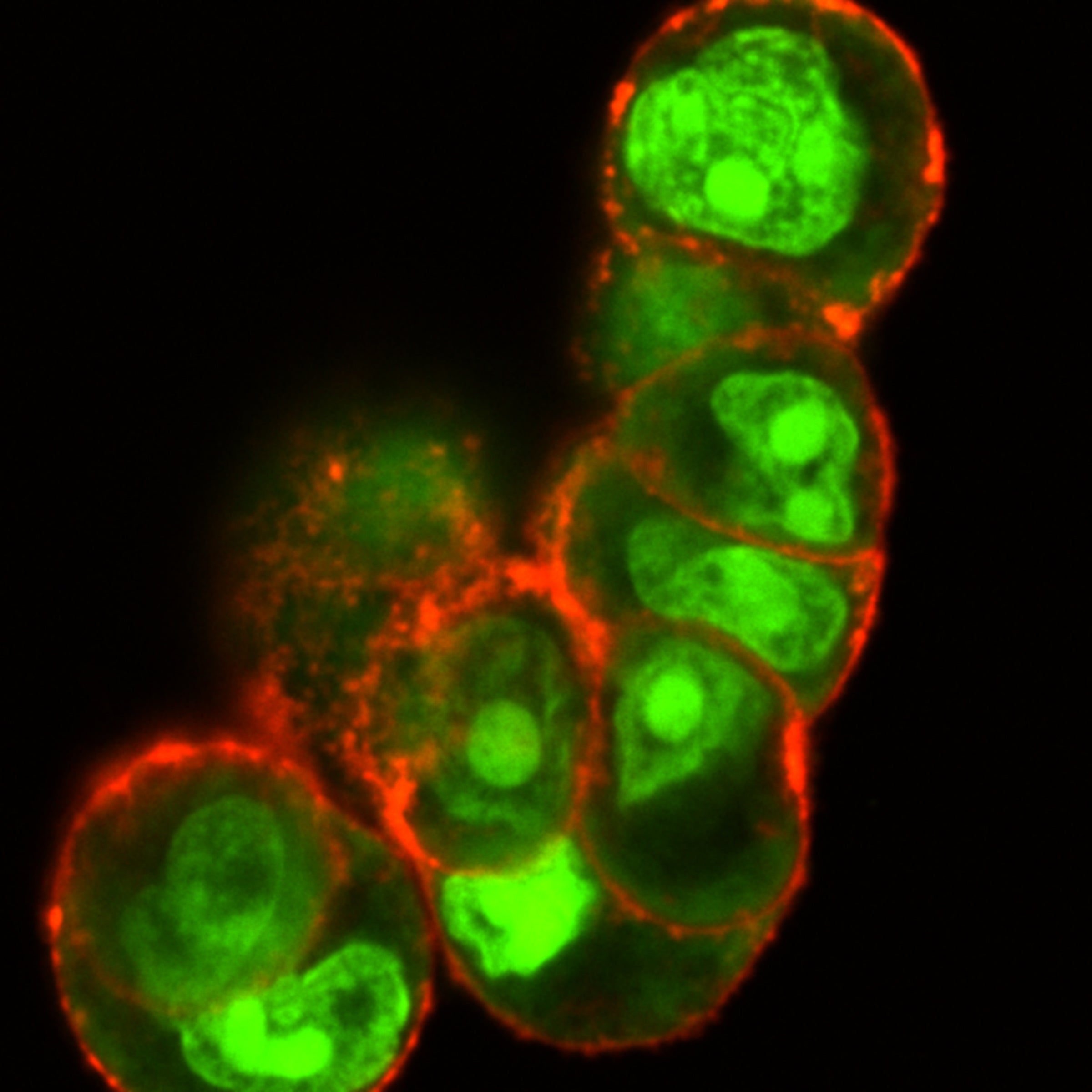
Soumyashree Sahoo, Chemistry Graduate Program
These colon cancer cells are being ruptured by reactive oxygen species that were generated inside the cancer’s own cells by a material synthesized by chemistry Ph.D. student Soumyashree Sahoo. As part of her research, Sahoo is working on synthesizing and delivering nanostructured therapeutics for cancer, in this case a protein-DNA conjugated nanostructure. This confocal laser scanning microscopy image reveals how the cell membrane (red) has been slightly ruptured, while the deadly reactive oxygen species (green) has been localized in the nucleus and along the cell membrane.
Visualizing Science Showcase