Scientists Study How DNA Repairs Itself Through Single Molecule Imaging
UT Austin scientists are doing research, which uses novel single-molecule imaging techniques partially developed by Finkelstein, and could lead to a better understanding of how cancerous cells repair their DNA.
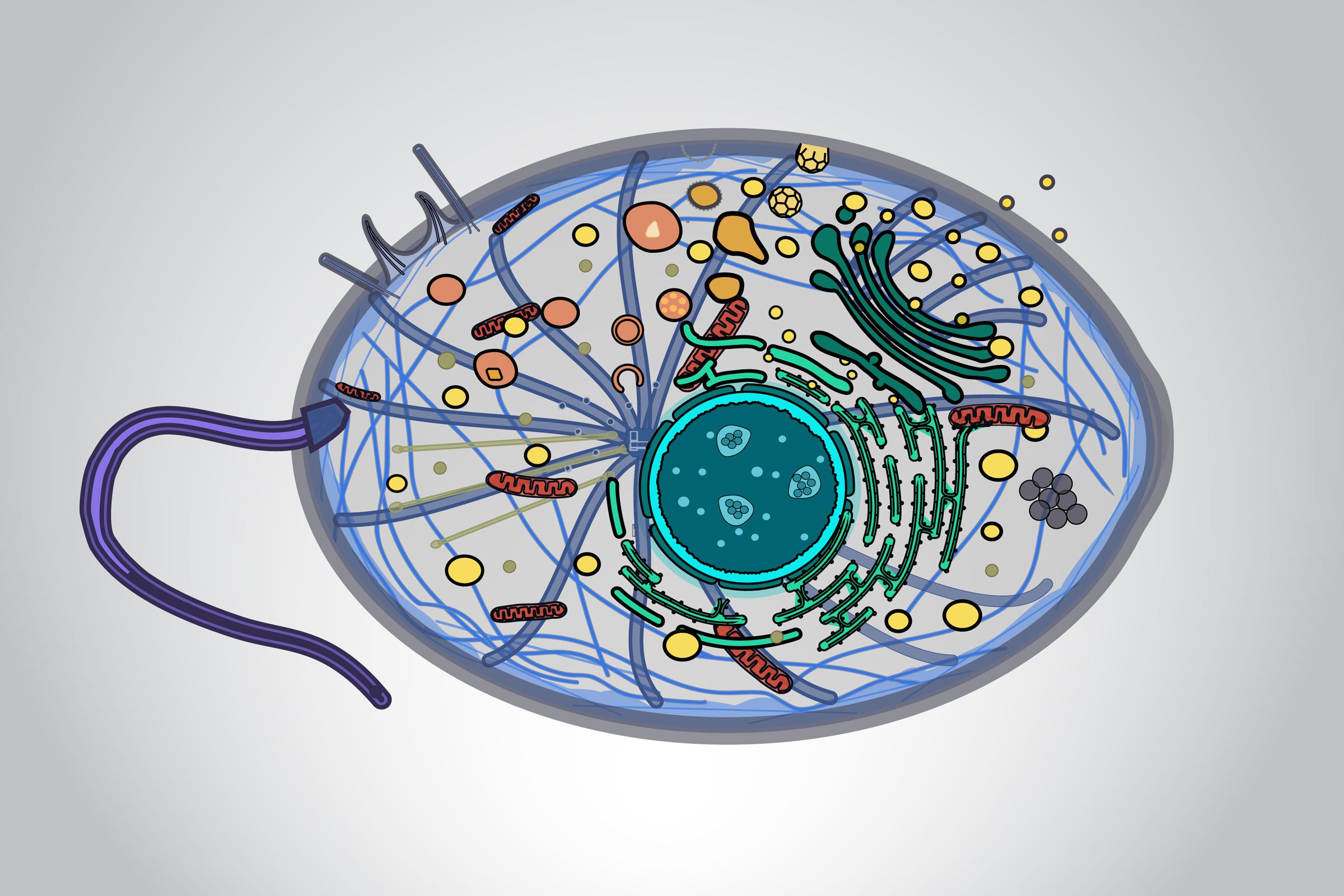
A group of scientists from UT Austin's Finkelstein Laboratory, headed by assistant professor in Molecular Biosciences Ilya Finkelstein, have imaged proteins which repair DNA and gained new insights into how the body regulates DNA repair. The research, which uses novel single-molecule imaging techniques partially developed by Finkelstein, could lead to a better understanding of how cancerous cells repair their DNA.
Understanding how a cancerous cell fixes its DNA is key in developing targeted genetic medicine that prevents the cell from ever fixing itself, eventually leading to the death of the cancer cell.
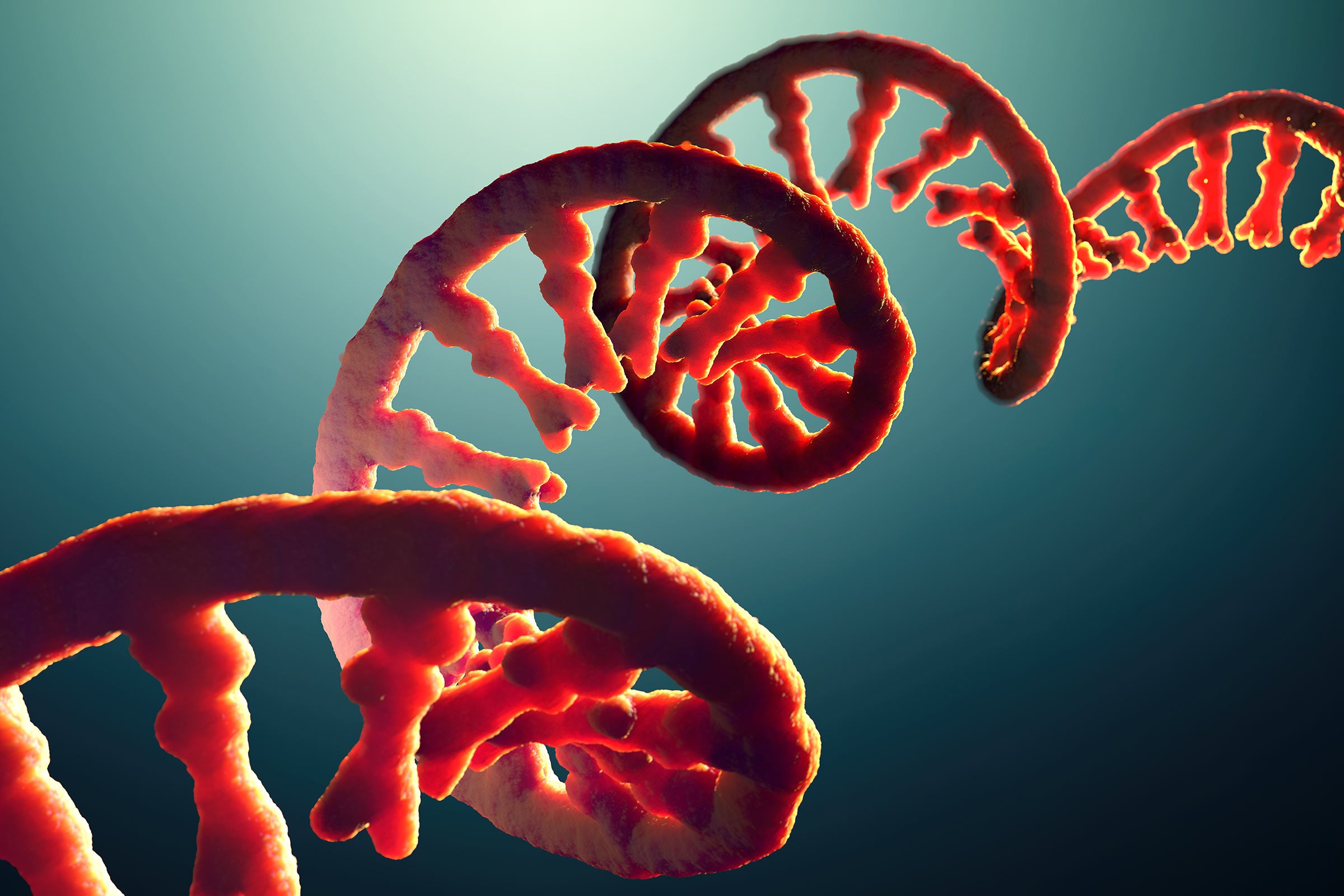
Our DNA is being constantly damaged in tiny ways, from things like the Sun's UV light, or from simple errors in replication. Because of this, the cell must have special enzymes to destroy damaged DNA and rebuild the segments that had been damaged. This problem of damaged DNA is even more acute in cancer cells because, as co-author and graduate research assistant Logan Myler explains, as "cancer cells are rapidly growing and mutating, they accumulate a lot of DNA damage." The 2015 Nobel Prize in Chemistry recognized the important role that understanding DNA repair plays in fighting cancer.
The research, published in the Proceedings of the National Academy of Sciences, focused on trying to understand Exo1, an enzyme which traverses one of the DNA's double helix strands and destroys the DNA it passes. Since such a destructive enzyme could be potentially dangerous in a cell, it's tightly regulated by another molecule, called Replication Protein A (RPA). The understanding of this enzyme, which is critical for the survival of all types of cancerous cells, might eventually lead to treatments that are effective against all cancers.
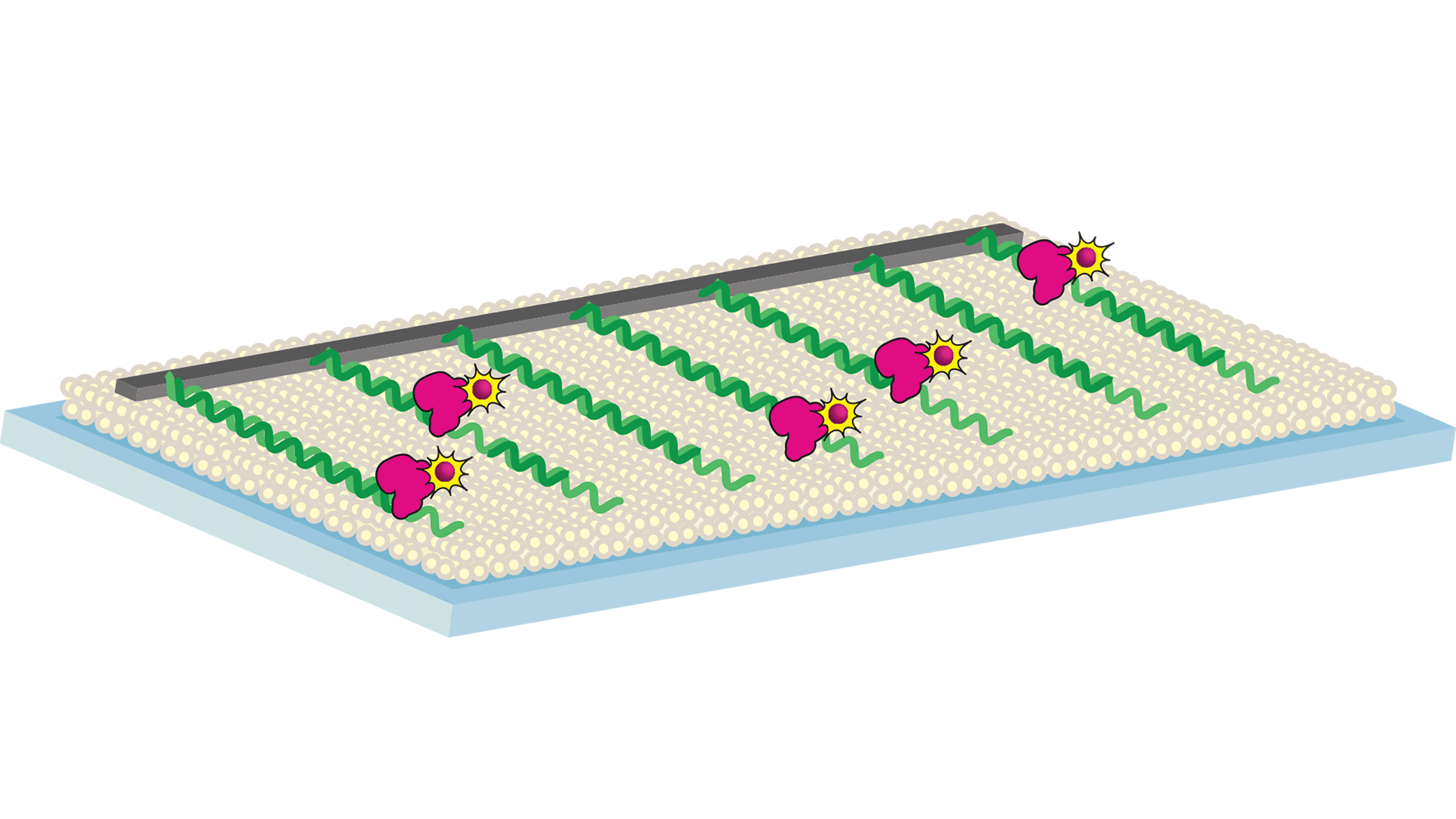
In order to accurately understand the DNA repair process, the team used a novel imaging technique which was able to see the process at the molecular level. The technique, which was developed at Columbia University with the help of Finkelstein, is called DNA Curtains. The process involves lining DNA up on a slide covered with material common in the cell and attaching the ends of the DNA to metallic edges. Images are taken from above and show the enzyme as it traverses the DNA.
Using the single-molecule imaging technique, the team aimed to clear up conflicting results from more traditional biochemical methods about how RPA worked. The previous results had not determined if RPA aided or inhibited the Exo1 enzyme. However, using the new imaging techniques the researchers were able to understand that RPA actually stopped Exo1 from destroying more DNA once it had reached the proper place in the strand.